Case of the Week #595
Prof at Akdeniz University Perinatology Department
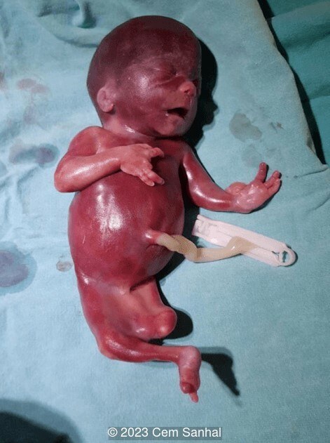
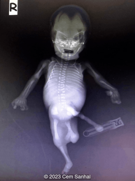
A 23-year-old G3P2 was referred to our clinic due to suspected multiple fetal anomalies at 23 weeks of gestation. Personal and family histories were unremarkable. There were no consangenious marriage and no drug use. We found oligohydroamnios and the following sonographic features. Conventional amniocentesis and G-karyotyping revealed normal findings. What is the diagnosis ?
View the Answer Hide the Answer
Answer
We present a case of Gollop Wolfgang Complex.
Our ultrasound demonstrated the following findings:
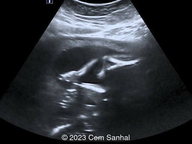
- Video 1-2 and Image 1: Distal femur duplication (bifid femur) with absence of lower extremity (tibia, fibula and foot) and ectrodactyly of hand
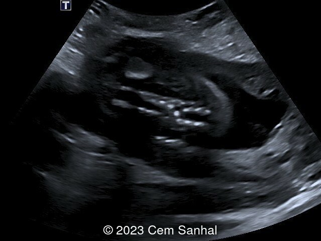
- Image 2: Hemivertebra
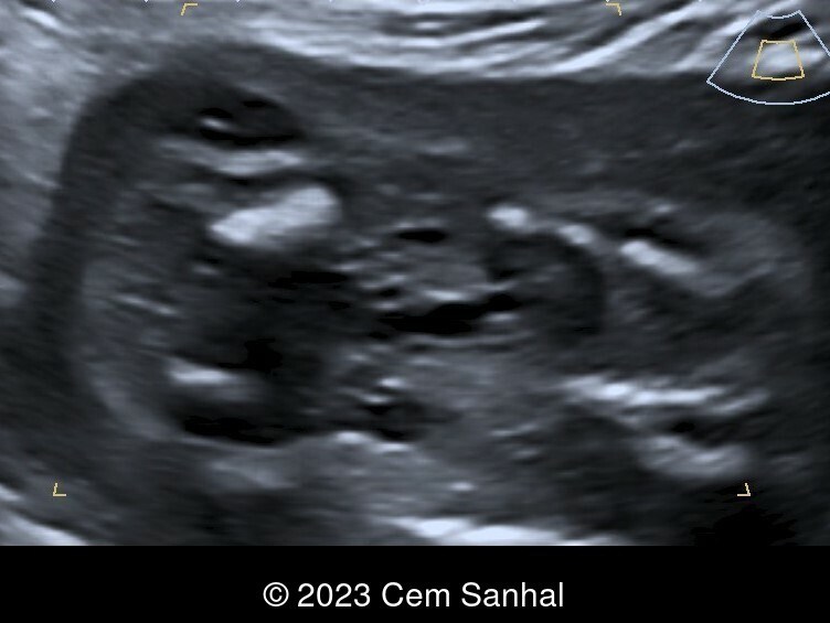
- Image 3: Multicystic dysplastic kidneys
Discussion
Gollop-Wolfgang complex is defined as the presence of a distal bifid femur (femur duplication) and tibial hemimelia with or without hand ectrodactyly [1,2]. Most cases of Gollop-Wolfgang complex are accompanied by other congenital defects, such as vertebral anomalies, anal atresia, cardiac malformations, tracheoesophageal fistula, esophageal atresia, renal anomalies, and limb defects, most commonly radial ray defects [3,4]. The incidence is reported to be 1 in 1,000,000 births [5].
Gollop-Wolfgang complex belongs to a skeletal dysplastic classification of dysostoses, which includes malformations or absence of individual bones singly or in combination [3]. According to the theory of Lewin and Opitz, the growth of the distal extremity is under the control of two developmental fields, the tibia and fibula. The tibial developmental field guides the development of the distal femur, tibia, and hallux. A defect in this field results in distal femur duplication, tibia agenesis, and preaxial polydactyly or ectrodactyly. The fibula developmental field controls the development of the fibula, lateral rays of the foot, lateral knee ligaments, proximal femur, acetabulum, and pubic bones. A defect in this field results in fibular hypoplasia, ectrodactyly, proximal focal femoral deficiency, and deficiency of lateral knee ligament. There is a strong association between the development of the fibula and tibial fields [3,6,7].
The etiology of Gollop-Wolfgang complex remains unknown. In two Japanese patients, a duplication and a triplication of a 210Kb chromosomal segment in 17p13.3 which included BHLHA9, was detected and considered a susceptibility factor for the limb malformation. Both autosomal dominant and autosomal recessive forms of inheritance were reported for Gollop Wolfgang complex [9].
Increased nuchal translucency between 11 and 14 weeks of gestation and tibial agenesis with femoral bifurcation, clubfoot, contracture of the knees, and agenesis of the metacarpal bones are the sonographic findings of Gollop Wolfgang complex [3]. Patients affected with this entity may also have ectrodactyly and cardiovascular or urogenital anomalies [8]. Amniocentesis can be performed for detection of chromosomal abnormalities, thus helping with genetic counseling.
References
[1] Ondari J, Kinyanjui J, Miano P, et al. Femoral bifurcation and bilateral tibial hemimelia: case report. Pan Afr Med J 2018;30:99
[2] Habou O, Magagi IA, Adamou H. Gollop-Wolfgang complex. J Neonatal Surg 2017;6(01):19.
[3] Vanderberg RH, Block T, Gates T, et al. Gollop-Wolfgang Complex: Clinical and Imaging Implications. Indian J Radiol Imaging 2021;31:721–724.
[4] Caforio L, Pagnotta G, Romiti A, et al. Prenatal diagnosis of Gollop-Wolfgang complex. Ultrasound Obstet Gynecol 2015;45(04):488–490.
[5] Pandy D, Pal MV, Nambiar J, et al. Gollop-Wolfgang complex-a rare limb deficiency syndrome: case report and review of literature. Internet J Gynecol Obstet 2007;9:1–5.
[6] Alessandri JL, Isidor B, David A, et al. Tibial developmental field defect in valproic acid embryopathy: report on three cases. Am J Med Genet A 2010;152A(11):2805–2809.
[7] Pavone L, Viljoen D, Ardito S, et al. Tworare developmental defects of the lower limbs with confirmation of the Lewin and Opitz hypothesis on the fibular and tibial developmental fields. Am J Med Genet 1989;33(02):161–164.
[8] Nlandu A, Docquier PL. Gollop-Wolfgang Complex : an alternative to amputation. Acta Orthop. Belg. 2013;79:239-242.
[9] Forzano F. Gollop-Wolfgang complex. Orphanet. https://www.orpha.net/consor/cgi-bin/OC_Exp.php?lng=en&Expert=1986. Last update 11/2019.
Discussion Board
Winners

Dianna Heidinger United States Sonographer

Javier Cortejoso Spain Physician

Mayank Chowdhury India Physician
Aysegul Ozel Turkey Physician
Halil Mesut Turkey Physician

Lech Dudarewicz Wallis and Futuna Physician

ALBANA CEREKJA Italy Physician

Hien Nguyen Van Viet Nam Physician
Ismail Guzelmansur Turkey Physician