Case of the Week #601
(1) Department of Genetics, Polish Mother's Memorial Hospital, Lodz, Poland; (2) St Mary's Medical Center, San Francisco, CA, USA
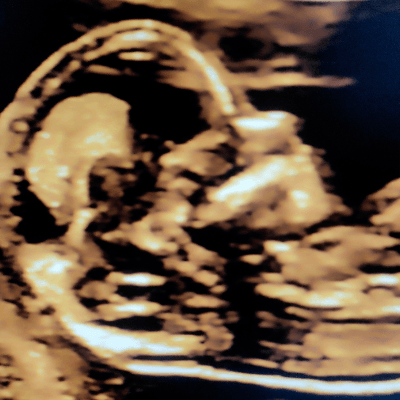
A pregnant woman presents at 13 weeks and underwent routine fetal scan presented in the video below. What is the finding?
View the Answer Hide the Answer
Answer
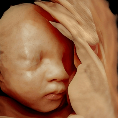
We present a case of cleft secondary palate in the first trimester diagnosed with "a superimposed line sign." Repeat ultrasound examination at 19 weeks confirmed the findings (Video 1).
Discussion
Cleft lip and cleft palate are common congenital anomalies with an incidence of approximately 1 to 2 in 1000 pregnancies [1-3]. Lack of fusion of the palatine shelves from the maxillary prominences starting at the 6th gestational week results in clefts of the secondary palate [3]. While cleft palate can be a challenge to diagnose prenatally [1], identifying a facial cleft in utero is important for patient counselling. Cleft palate is associated with other congenital malformations in 23-82% of cases and with recognized syndromes or chromosomal anomalies in 7-27% [1-3]. In affected families, the recurrence rate is 3.5% for first degree relatives, though can be as high as 4.6% in cases with more severe anatomical defects [4].
Diagnosis of a cleft palate is challenging as its dome shape and acoustic shadowing from the alveolar ridge of the maxilla make it difficult to visualize on ultrasound. In normal fetuses, the midsagittal view of the secondary palate shows a double echogenic line: the vomer bone is represented by the shorter, more cranial line while the hard secondary palate is a longer, more caudal line. Together they comprise the maxillary line [3]. These detailed anatomic landmarks were described by Rotten et al in 2004 in their sonographic assessment of the fetal face [5]. In a fetus with a cleft of the secondary palate, the line formed by the palate is absent, thus only the vomer bone is visualized. The result is a single line instead of the characteristic superimposed double line. The “superimposed line sign” is best visualized between 13 and 17 weeks of gestation. In a study reviewing nearly 10,000 pregnancies, the superimposed line sign had a sensitivity of 89.5% in detecting a cleft of the secondary palate, a specificity of 99.8%, a positive predictive value of 43.6% and a negative predictive value of 99.9% [3].
Other ultrasonographic signs that have been studied include the maxillary gap, which can also be seen in the midsagittal view of the fetal face [6]. In a case-control study including 86 fetuses with cleft lip and/or palate, a maxillary gap was observed in 7% of controls, 65% of fetuses with isolated cleft lip and/or palate and 96% of fetuses with facial cleft associated with other anomalies. The size of the maxillary gap in controls was <1.5mm. During the 11 to 13 week ultrasound examination, some parts of the maxilla may still show lack of ossification, which may explain the gap in normal controls. In contrast, the maxillary gap was between 1.5 and 5.0mm in over half of the fetuses with isolated cleft lip and/or palate. Thus, a small maxillary gap in the midsagittal view in the presence of an intact maxilla in the coronal view can be considered a normal finding, while a larger gap may be suggestive of a facial cleft [6].
Several three-dimensional techniques have been proposed for imaging the secondary palate [7], though 3D sonography requires adequate volume acquisition with the fetal neck in slight extension and the presence of fluid between the fetal tongue and the palate [3]. The retronasal triangle view describes visualization of the primary palate and frontal processes of the maxilla in the coronal plane of the fetal face [7]. A facial cleft may be present if the retronasal triangle appears distorted or if the base of the triangle is incomplete. In a study of 240 pregnancies, the sensitivity and specificity of the retronasal triangle view for detecting facial clefts was 85% and 93%, respectively [7].
In conclusion, indirect signs of a cleft palate in the first trimester are important for identification of this common congenital malformation. In larger clefts the maxillary gap can be visible, while in smaller clefts, the lack of a superimposed line sign may lead to the diagnosis, as in our case. If either sign is present, additional detailed views of the fetal face and 3D sonography may aid in the diagnosis. Given the strong association of cleft palate with structural anomalies and genetic syndromes, referral to a tertiary center where molecular diagnostics can be performed is recommended.
References
[1] Offerdal K, Jebens N, Syvertsen T, et al. Prenatal ultrasound detection of facial clefts: a prospective study of 49,314 deliveries in a non-selected population in Norway. Ultrasound Obstet Gynecol. 2008 Jun;31(6):639-46.
[2] IPDTOC Working Group. Prevalence at birth of cleft lip with or without cleft palate: data from the International Perinatal Database of Typical Oral Clefts (IPDTOC). Cleft Palate Craniofac J. 2011 Jan;48(1):66-81.
[3] Lakshmy SR, Rose N, Masilamani P, et al. Absent 'superimposed-line' sign: novel marker in early diagnosis of cleft of fetal secondary palate. Ultrasound Obstet Gynecol. 2020 Dec;56(6):906-915.
[4] Grosen D, Chevrier C, Skytthe A, et al. A cohort study of recurrence patterns among more than 54,000 relatives of oral cleft cases in Denmark: support for the multifactorial threshold model of inheritance. J Med Genet. 2010 Mar; 47(3): 162–168.
[5] Rotten D, Levaillant JM. Two- and three-dimensional sonographic assessment of the fetal face. 1. A systematic analysis of the normal face. Ultrasound Obstet Gynecol. 2004 Mar;23(3):224-31.
[6] Chaoui R, Orosz G, Heling KS, et al. Maxillary gap at 11-13 weeks' gestation: marker of cleft lip and palate. Ultrasound Obstet Gynecol. 2015 Dec;46(6):665-9
[7] Martinez-Ten P, Adiego B, Illescas T. First-trimester diagnosis of cleft lip and palate using three-dimensional ultrasound. Ultrasound Obstet Gynecol. 2012 Jul;40(1):40-6.
Discussion Board
Winners

Javier Cortejoso Spain Physician
Ana Ferrero Spain Physician

Ivan Ivanov Russian Federation Physician
Boujemaa Oueslati Tunisia Physician

Annette Reuss Germany Physician

doğa öcal Turkey Physician

ALBANA CEREKJA Italy Physician
Deval Shah India Physician
Murat Cagan Turkey Physician
Ionut Valcea Romania Physician
reyhan ayaz Turkey Physician
Subapriya Kandasamy India Physician
Anette Beverdam Netherlands Sonographer
Vandana Yakub India Physician
Shina Kaur India Physician
Aylin Özgün Turkey Physician
Lê Đức Viet Nam Physician
Costin Radu Lucian Romania Physician
Gaurav Sharma India Physician
Alpana Watwe India Physician