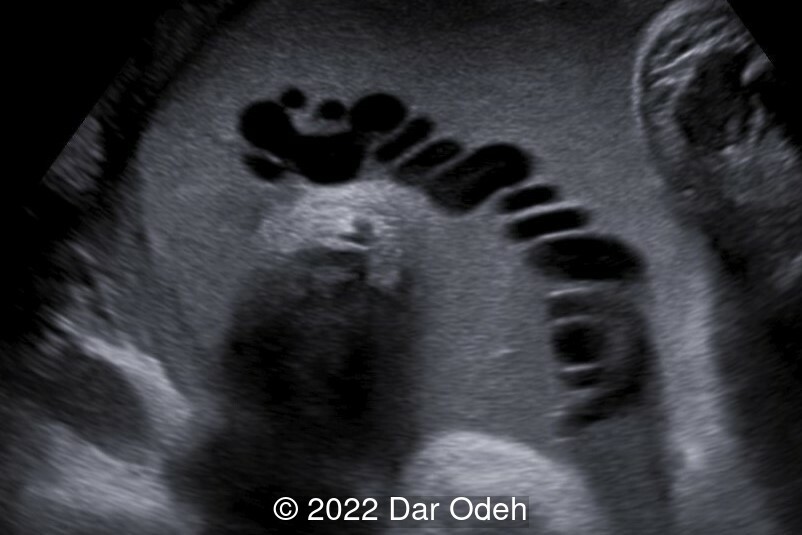
Highly echogenic amniotic fluid
Omayyah Dar-Odeh*, Samih Abdel-Jawad**, Rua'a Abdel-Jawad***, Raghad Abdel-Jawad****, Rahaf Abdel-Jawad******Private sector in Amman- Jordan, **Private sector in Amman- Jordan,***King Hussein Cancer center in Amman- Jordan, ****Hashemite University in Jordan, *****Hashemite University
Article Published: Oct 17, 2022
A 39-year-old G3P2 woman, was referred at 39 weeks gestation due to undefined pathology. The patient reported an uneventful course of pregnancy, however, no nuchal translucency scan, nor detailed anatomy scan were done previously. Our ultrasound examination revealed the following findings:
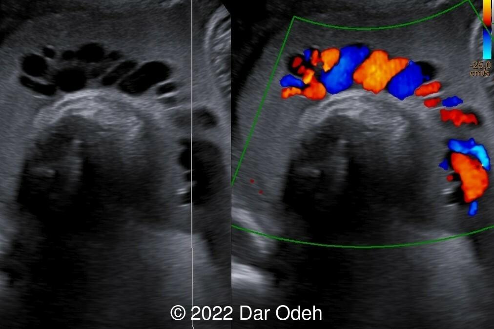
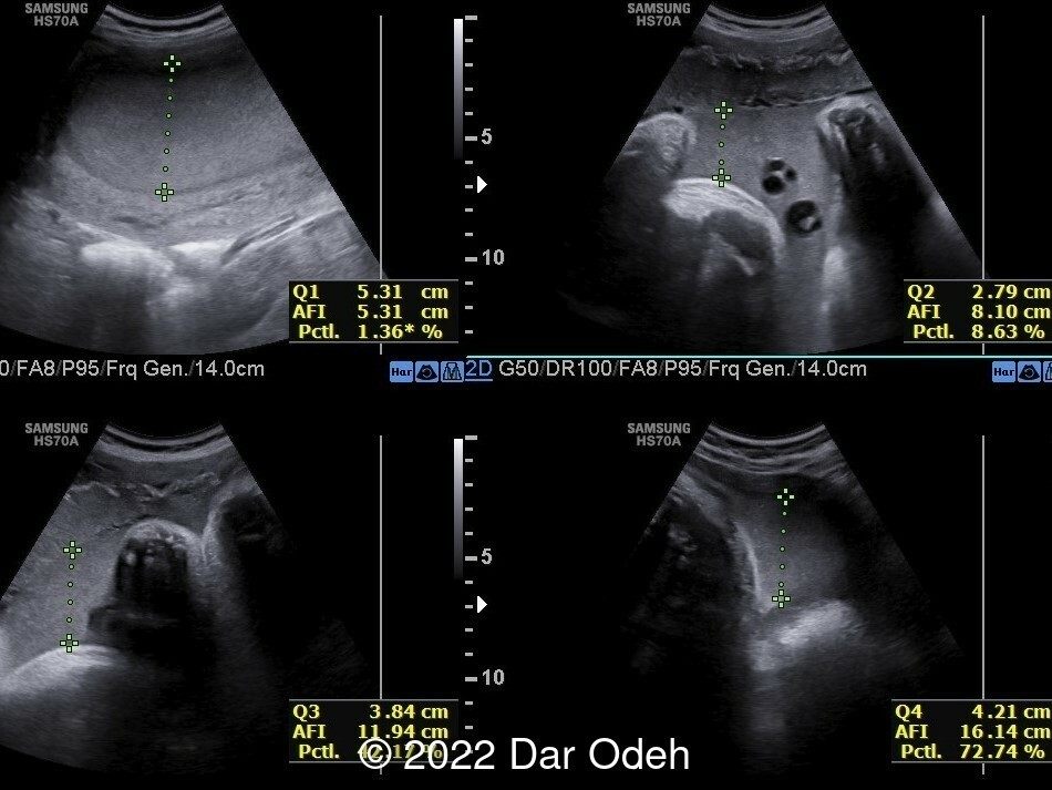
Our ultrasound findings revealed highly echogenic amniotic fluid. We advised conservative management and counselled the couple to wait until labor pains start. Biophysical profile with nonstress test were reassuring. However, due to clinicians' concerns, delivery was induced at 39 weeks +3 days with careful fetal monitoring. A healthy male baby weighing 3840g was delivered vaginally after 6 hours of induction with prostaglandin and oxytocin. Amniotic fluid was found to be clear at time of delivery.
Discussion
Amniotic fluid is the liquid that surrounds a developing fetus in the amniotic sac and is usually clear to pale yellow in color. Amniotic fluid composition is complex with many maternal and fetal constituents. The composition of amniotic fluid changes with the gestational age with an average pH of 7.2 and specific gravity of 1.0069–1.008. Echogenicity of amniotic fluid indirectly represents the size, number, and distribution of particles in the fluid and in turn turbidity of amniotic fluid. This could give rise to the detection of echogenic particles, visualized as homogeneously echogenic amniotic fluid on ultrasound, and known as amniotic fluid "sludge".
Amniotic fluid sludge is dense aggregates of particulate matter. The presence of echogenic amniotic fluid at term gestation on sonography is uncommon, with an incidence at term of 3.2 %. Echogenic amniotic fluid creates a dilemma in the mind of the clinician. Very echogenic amniotic fluid has been variably attributed to meconium, blood, or vernix caseosa. Many studies have shown that the presence of meconium is unlikely. However, most reports are case reports, and are not been investigated by amniocentesis. Very echogenic amniotic fluid in the third trimester is most often due to vernix caseosa, which is a complex fatty substance derived from the desquamated epithelial cells and sebaceous material. This sonographic finding is not a reliable indicator of meconium or blood in amniotic fluid and should not typically alter antenatal management.
Congenital conditions associated with particulate matter in the amniotic fluid include harlequin ichthyosis and epidermolysis bullosa letalis.
References:
1. Brown, D L et al. “Very echogenic amniotic fluid: ultrasonography-amniocentesis correlation.” Journal of ultrasound in medicine : official journal of the American Institute of Ultrasound in Medicine vol. 13,2 (1994): 95-7.
2. Buyuk, Gul Nihal et al. “Echogenic particles in the amniotic fluid of term low-risk pregnant women: does it have a clinical significance?.” Journal of obstetrics and gynaecology : the journal of the Institute of Obstetrics and Gynaecology vol. 41,7 (2021): 1048-1052.
3. Karamustafaoglu Balci, Burcin, and Gokhan Goynumer. “Incidence of echogenic amniotic fluid at term pregnancy and its association with meconium.” Archives of gynecology and obstetrics vol. 297,4 (2018): 915-918.
4. Kaluarachchi, Athula et al. “Hyperechoic amniotic fluid in a term pregnancy.” Journal of family medicine and primary care vol. 7,3 (2018): 635-637.
5. Hill, L M, and R Breckle. “Vernix in amniotic fluid: sonographic detection.” Radiology vol. 158,1 (1986): 80.